fbpx
Skip to main content

Laser Collaboration, 33 Partners from Around the Globe Represented ~ Erchonia 2018

World’s Laser Therapy Advocates Convene in Orlando, FL, USA on May 3rd-4th, 2018

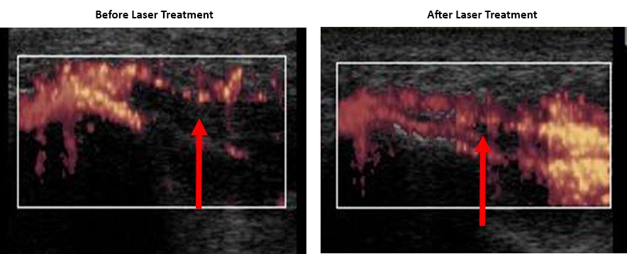
before and after laser treatment


MELBOURNE, FLA. (PRWEB) MAY 01, 2018 We are verging on a global precipice: the increase in the number of debilitating brain diseases is growing exponentially, obesity rates are skyrocketing, more than ever before, patients who experience pain are becoming addicted to opioids. Traditional medicine is on the brink of a revolution in the healing arts; this revolution is called low-level laser therapy (3LT®)!  Florida based low-level laser technology manufacturer Erchonia Corporation will host their annual Partner’s Meeting at the Hyatt Regency Orlando on May 3rd-4th, 2018.  With over 2 decades of published laser research and 15 FDA 510(k)s on laser therapy leading up to this conference, the advancements in medicine and laser therapy will impress attendees and international partners alike.

World renowned researchers and scientists will prepare substantiated lectures reviewing existing low-level laser FDA market clearances and disclose (3) new, Level (I) clinical trials, their applications and the long-term developments for low-level laser therapy.  The new laser application line-up includes brain diseases, low back pain and peripheral neuropathy just to name a few.

“We are very excited by the growth that we are achieving across the world.  We are confident Erchonia Lasers will continue to prove to be ‘THE’ prominent choice for those seeking laser therapy.  The impeccable presenter line-up we have scheduled is like none other.” -Charlie Shanks, VP of Erchonia Corp

Researchers, International Partners and Erchonia Laser Enthusiasts are invited to share in a goldmine for laser therapy.  The educational meeting will be followed by a cocktail reception at The Red Coconut Club at Universal City Walk where attendees will meet the presenters from around the globe.

*New indications discussed during this event are going through FDA clinical trials.  Go to www.ClinicalTrials.gov, search “Erchonia”.  Devices for new indications have not been FDA market cleared.  New devices will not be introduced to market until FDA 510(k) is obtained.

Founded in 1996, Erchonia Corporation is the world leader in low-level laser technology. The company created the low-level laser category in 2002 when it received an FDA 510(k) market clearance for low-level lasers. Erchonia was the first company to receive this FDA 510(k) distinction. For more information, visit www.erchonia.com or call 888-242-0571.

For additional information, image and interview requests, contact Erchonia Corporation (888) 242-0571.

Related Links https://www.Erchonia.com


https://www.prweb.com/releases/2018/05/prweb15452126.htm

Low Level Laser Therapy for 360°- Degree Body Sculpting

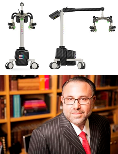
Michael H. Gold, M.D., FAAD from Gold Skin Care Center presenting Thursday 5th April @ 10:00AM at AWMC 2018 ~ Monaco.


Dr. Michael H. Gold presenting the Verjú laser device

Verjú LLLT by Erchonia, FDA Cleared for circumferential reduction of the waist, hips and thighs and temporary reduction in the appearance of cellulite will be featured on the programme at the Aesthetic and Anti-Aging Medicine Congress 2018.  The Verjú laser produces a low-level output that has no thermal effect on the body’s tissue.  Its unique technology does not kill or damage fat cells, but instead creates a transitory pore in each cell for the fat to leak out, which is then processed naturally through the lymphatic system, leaving the cell intact for important endocrine function.

The 510(k) is a premarketing submission to the FDA that demonstrates that the device marketed is safe and effective. The premarket approval for the 510(k) is the most rigorous type of device marketing application accepted by the FDA.  The Verjú Laser System obtained its 1st 510(k) clearance in 2012.

Verjú Study Results – NO lifestyle changes – Published in American Journal of Cosmetic Surgery for Body Contouring and Lasers in Surgery in Medicine for Appearance of Cellulite.  There were no adverse events in either study.

A world leading Dermatologist and Cosmetic Surgeon, Michael H. Gold, M.D., FAAD, will present Thursday 5th April @ 10:00AM.  Dr. Gold’s message is ‘A Better You’!

Simon Ramshaw, Managing Director of Erchonia Lasers LTD. comments, “Having an expert such as Dr. Gold recognize the quality of our research and the clinical utility of Erchonia’s Verjú Laser

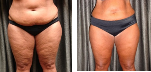
Before and after photos of successful laser therapy for obesity

System is truly an honor.  Quality clinical research is pivotal for our overall success.  Erchonia’s commitment to research stands out in aesthetic medicine.”

Founded in 1996, Erchonia Corporation is the world leader in low-level laser technology. The company created the low-level laser category in 2002 when it received an FDA 510(k) market clearance for low-level lasers. Erchonia was the first company to receive this FDA 510(k) distinction. For more information, visit www.erchonia.com or call 888-242-0571.

For additional information, image and interview requests, contact Erchonia Lasers Ltd at +44(0)1491 821135.

Related Links https://www.Erchonia.com


WHAT STUDIES SAY ABOUT CLASS IV LASERS & SPECIFICALLY, THEIR IMPACT ON CANCER CELLS


 class IV lasers' impact on cancer cells
The National Cancer Institute (NCI) reports that 39.6 percent of all Americans will receive a cancer diagnosis at some point in their lives. The NCI goes on to explain that cancer is one of the top causes of death across the globe, with new diagnoses expected to increase twofold-from 14 million to 22 million-within the next twenty years. Because of statistics like these, many researchers are working diligently to learn more about cancer cells, as well as to discover what makes them form and grow. One area of study they’ve spent some time in recently involves the use of medical lasers. Class IV lasers, specifically.

Lasers and cancer cell research

One such study was published in the American Academy of Physical Medicine and Rehabilitation’s PM&R Journal in November of 2017. This particular piece of research involved the use of two different types of cells: saos-2 osteoblast-like osteosarcoma cells and A549 human lung carcinoma cells.

After preparing the culture plates, each one was subjected to laser irradiation either one, two, or three times with d:YAG lasers, the power output ranging from 0.5 to 3 watts. Approximately 24 hours after receiving the last laser application, the plates were analyzed to see what change, if any, existed to the cells.

It was during this analysis that researchers discovered that, the more laser applications a specific culture plate received, the higher the proliferation rate, or the more these cancerous cells increased in number, when compared to control plates. Thus, they concluded that this type of laser therapy “could activate precancerous cells or increase existing cancerous tissue.”

Other studies have found similar results. For instance, in November of 2009, BMC Cancer published a study involving mice with melanoma cells. For purposes of this particular study, some of the mice served as the control, some were exposed to three sessions of irradiation lasting 60 seconds each (dose 150 J/cm2), at the rate of one per day for three days, and the rest were exposed to three sessions of irradiation lasting 420 seconds (dose 1050 J/cm2).

Upon conclusion of the final session, each mouse was analyzed to see if there were changes in the melanoma cells. The group that was exposed to 60 seconds of lower level laser therapy (150 J/cm2) showed results “not significantly
different from the controls.”

However, the same could not be said for the other mice, the ones exposed to the more powerful laser doses (1050 J/cm2), as the researchers reported that they experienced “significant increases in tumor volume, blood vessels and cell abnormalities compared to other groups.


So, are medical lasers safe?

Findings like these underline the importance of using lasers with the appropriate strength and within the appropriate
class to obtain the desired medical result without risking the patient’s health and safety.

This involves using lasers within the lower classifications (Class I, 11, or Ill) as many of these have been found safe for enhancing health and wellness without impacting a person’s risk of cancer or increasing their cancerous cell activity. It also means using lasers from an FDA approved company, thus  protecting yourself as well as your patients.

Class IV medical lasers defined: cancer cell

The U.S. Food and Drug Administration (FDA) adds to the topic by sharing that lasers can be placed in one of four hazard classes, ranging from Class I to Class IV, with some of these classes containing subclasses within them. However, For simplicity’s sake, the FDA says that the best way to understand the differences is, “the higher the class, the more powerful the laser is and the greater the potential to pose serious injury if used improperly.” Because these are the highest level of lasers, this causes many to question what impact they may have on the human body. Specifically, some researchers have set out to discover what effect they may have on cancerous and precancerous cells.


Sources
1. The National Cancer Institute – https://www.cancer.gov/about-cancer/understanding/statistics

2. American Academy of Physical Medicine and Rehabilitation’s PM&R Journal in November of 2017 – https://www.ncbi.nlm.nih.gov/pubmed/29160001

3. BMC Cancer – https://bmccancer.biomedcentral.com/articles/10.1186/1471-2407-9-404

4. U.S. Food and Drug Administration (FDA) – https://www.fda.gov/Radiation-EmittingProducts/RadiationEmittingProductsandProcedures/HomeBusinessandEntertainment/LaserProductsandInstruments/default.htm

Disclaimer
*The material presented is being made available for educational purposes only. This article was compiled based on research collected through online and medical publications. Erchonia is not liable for any use you may make of such information. The views are presented to inform readers of possible dangers of high powered lasers. All of the opinions presented do not present any medical claims not currently stated through government agencies. The approaches, information and opinions may not be applicable to all cases, and each health care provider must use their own independent medical judgment in treating any individual patient or condition.


5 Myths of Laser Body Contouring

Like most other popular weight loss procedures, laser body contouring has its share of misconceptions. In an effort to clear up some of those myths, we’ll look at five of the most common and give you the truth of the matter.

  1. Laser body contouring is too expensive.

    When compared to traditional surgical liposuction, which runs anywhere from $1,500 to $7,500, the cost of laser body contouring isn’t so bad after all at $1,000 to $2,500.happy woman at beach

  2. Laser body contouring is painful.


    With most patients having laser body contouring treatments in their plastic surgeon’s office and then going on about their day, this myth is easily discounted. Laser contouring doesn’t require any kind of anesthesia—in fact, most patients say it feels like nothing or like a flashlight shining on them. When compared to traditional inpatient liposuction with all its incisions, bruising, and swelling, laser body contouring’s noninvasive techniques are much more easily tolerated.

  3. Laser body contouring takes a long time.

    Each session typically lasts 30 to 40 minutes. Each treatment typically involves 3 sessions per week over two weeks. That’s 6 sessions at 30 to 40 minutes each. The average American spends about the same amount of time watching television each weekend. You decide.

  4. Laser body contouring is dangerous.

    Unlike traditional liposuction, laser body contouring carries few risks. There are no incisions, no need for anesthesia, and no cause for antibiotics. There is no scarring involved. Laser body contouring is as safe as it gets in medical procedures—with the right tools, the right training, and the right hands on the tools, you have nothing to worry about.

  5. Laser body contouring doesn’t work.

    From the number of scientific studies to the number of happy, satisfied customers, it’s easy to see that this one is just plain wrong. Laser body contouring has been around for many years, and the positive results from patients can’t be denied. How well the treatment lasts depends on the patient’s willingness to watch diet and exercise and not allow themselves to pack the pounds back on. But the treatments in and of themselves are highly effective at helping patients lose unwanted fat deposits.

While laser body contouring may not be for everyone, there’s no need to believe all the false myths and misconceptions about it. Knowing the facts can help you make a better informed and educated decision. If you would like to learn more about laser body contouring, check out our My Zerona laser scanner.

How 3LT Can Help Our Equine Friends

Humans domesticated horses millennia ago, changing the way our ancestors traveled, fought, and survived. They are majestic, beautiful creatures built for speed and power. 

Sadly, one of the most serious and devastating diseases affecting horses, ponies, and other equine animals is laminitis. Let’s take a closer look at laminitis and how low-level laser therapy can give our equine companions a leg up.

What is Laminitis?

Laminitis describes a condition wherein the laminae—the tissues bonding the hoof wall to the pedal bone in a horse’s hoof—become weakened and inflamed from disruptions in blood flow, leading to tears in the structure supporting the pedal bone within the hoof. Laminitis typically occurs in a horse’s front feet. The condition is caused by various physical and metabolic issues, including:

  • An excessive intake of grain or grass
  • High levels of insulin
  • Enlargement of the pars intermedia in the pituitary gland
  • Impact from riding on hard surfaces
  • Stress from long distance travel

This can result in tremendous pain, lameness, and deterioration in the hoof. Left untreated, laminitis can cause the pedal bone to rotate and point downwards. In worst cases, the pedal bone will penetrate through the hoof wall.

Laminitis greatly reduces a horse’s usefulness, and many horse owners are forced to put down the horse to prevent further suffering.

Treating Laminitis

Many traditional treatments are expensive and time-consuming and don’t guarantee full recovery. These include changing your horse’s diet, providing greater hoof care, and moving your horse to a different enclosure featuring deep shavings or sand. Severe cases wherein the pedal bone has sunken through the hoof require surgical procedures involving tendon release, but this can put the horse at risk of infection or cause damage to surrounding structures.

Low-level laser therapy has been used in humans to treat joint pain, edema, soreness, and wounds, but veterinarians have extended these laser treatments to horses suffering from laminitis. Studies show that the photon energy in a low-level laser stimulates blood vessels in a horse’s foot, promoting greater circulation, better tissue nutrition, and ultimately faster healing. Laser therapy also greatly reduces the chance of infection or damage to surrounding areas as the procedure is entirely non-invasive.

Animal Health Options, a purveyor of innovative and effective supplements for animal wellness since 1990, has found success in incorporating low-level laser therapy into its treatment for laminitis. Horses undergo low-level laser sessions two to three times a week. This is coupled with:

  • A restrictive diet to reduce weight and make up for insulin resistance
  • Plenty of lying down to keep pressure off the affected feet
  • Visits with a farrier to trim hooves to correct the angle of the feet

Preventing Laminitis

One of the best ways to treat laminitis is to prevent it from happening altogether. While you can’t always predict your horse’s health, you can control parts of his environment, primarily his diet. Too much grain or lush green grass leads to excessive sugars stored in the hind gut. When these sugars are absorbed, the horse develops hyperinsulinemia (an overload of insulin), which can trigger laminitis. A bad diet can also lead to obesity, putting more pressure on your horse’s hooves. To keep your horse’s diet in check:

  • Feed your horse a high fiber, forage-based diet, comprising a mixture of mature grass, hay, and alfalfa. Vegetable oils can be included in this diet for added calories.
  • Carefully manage grazing. Considering grazing your horse at night, when sugar levels are lowest.
  • Avoid hard feed unless your horses are performing hard work.

Maintain a regular hoof trimming schedule for good hoof health. Your horse may also need specialist shoeing for proper support.

Zerona Laser Fat Attack – Muscular Development Magazine May 2010 Issue

Topical Fat-Buster:  Lose 7 Inches of Fat in 20 Minutes — No Bull!
By Dan Gwartney, MD

muscular development cover

Zerona Laser in Fitness RX for Men May 2010 Issue

Topical Fat-Buster:  Lose 7 Inches of Fat in 20 Minutes-NO BULL
By Dan Gwartney, MD

Fitness RX Men Cover

Zerona Laser in Fitness RX for Woman June 2010 Issue

Topical Fat-Buster:  Lose 7 Inches of Fat in 20 Minutes-Really!?!
By Dan Gwartney, MD
Fitness RX Women Cover

BIOMODULATION EFFECTS ON CELL MITOSIS AFTER LASER IRRADIATION USING DIFFERENT WAVELENGTHS

R. Sroka, C. Fuchs, M. Schaffer, U. Schrader-Reichardt, M. Busch, T. Pongratz, R. Baumgartner.

LFL Laser – Research Laboratory – Clinic of Urology and Clinic of Radiotherapy, University Munic, FRG

The biostimulative effects on cell mitosis induced by laser light at different wavelengths in cell cultures had been investigated, Murine skeletal fibroblasts (C2), normal urothelial cells (HCV29), human squamous carcinoma cell line of the mouth (ZMK) and urothelial carcinoma cells (J82) were irradiated with laser light at ^=488, 630, 640 and 805+25pm using a computer controlled irradiation chamber. The irradiance was set to 10mW/cm(2) and 100mW/cm(2), while the irradiation varied between 2 and 201/cm(2). The mitotic was determined by single cell counting after Orecein staining 24h post irradiation.

The mitotic rate showed a wavelength dependency with maxima at ^=635 and 805+nm for HCV29 and J82 cells. While the mitotic rate of C2 and J82 cells has the maximum value at about 41/cm(2), the maximum was at about 81/cm(2). ZMK cells showed no increase. At ^=805+25pm C2 and ZMK cells showed slight decrease in the mitotic rate after irradiation with 201/cm(2). An irradiation of 10mW/cm(2) was more effective than with 100m/Wcm(2). The biostimulation of the mitotic rate of both normal and tumor cells depends on the wavelength, irradiation and irradiance and on the cell line. The wave length dependency in the ^=630 to 640nm range could indicate a participation of endogenous porphyrins. Because the results show stimulative as well as inhibiting effects it should be considered to change the term biostimulation “into biomodulation.”

Information Application: 
Supports laser induced biomodulation

Would Albert Einstein use an Erchonia handheld laser?

ChiroEco

Background

The progression of low-level laser therapy as a viable therapy has made tremendous strides since the early investigations conducted nearly a century ago to assess how light can affect atom bound electrons. The photoelectric effect, a theory postulated by Max Plank and later proven by Albert Einstein in the early 1900s, identified that light was quantized and carried the force for the electromagnetic field, acting both as a wave and particle.

It was described by Einstein that light of a particular energy or color, as described by the following equation (Energy = plank’s constant x speed of light/wavelength), is capable of inducing electron emission at higher frequencies (lower wavelength) and independent of the intensity of light, an occurrence known as ionization (Fig. 1).
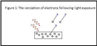
ionization of electrons following light exposure

Einstein revealed that no matter how high the intensity was increased, if the photon did not possess a specific energy (frequency), the electron could not be ionized. Adjusting to the wavelength capable of ionization and increasing the intensity did, however, promote a greater number of photonic collisions with the atoms thus escalating the number of electrons emitted from the atom. This simple concept of increasing the intensity to obtain enhanced photoreactivity however does not translate well when applied to the human model.

It has been more than 100 years since Einstein stimulated metal surfaces to demonstrate the photoelectric effect and since that time research has demonstrated that electrons when stimulated with the correct wavelength can become excited, leaving the ground state and entering an excited state, becoming more bio-reactive as electrons can temporarily reside on the outer orbital of an atom.

A key word in the previous sentence is “temporarily,” as I am reminded of the phrase “what goes up, must come down”; and in fact, the electron must return to a ground state, and in doing so, energy is released.

The release of energy can result in transient heating of the photoabsorbing molecules within nonphotosynthetic cells, and too much electron excitation induces large quantities of heat release. Identified photoabsorbing complexes are simple proteins that possess prosthetic metal groups forming chromophore structures, and like all other proteins, function under ideal parameters regarding temperature and pH is essential. Increasing the intensity may sound like a clever means to enhance photobiomodulation of cells, but recent clinical trials have demonstrated the opposite.

Research

A recent paper “Biphasic Dose Response in Low Level Light Therapy” co-authored by Dr. Michael Hamblin, Professor at Harvard Medical School, was published in the journal Dosage in 2009.1 Dr. Hamblin and colleagues discussed the dogma of intensity and dosage as they apply to laser therapy.

The authors assessed numerous clinical investigations and concluded that the delivery of lower intensity across greater treatment times yielded higher utility.1 Dr. Hamblin et al. postulated that higher intensity devices generate a detrimental level of reactive oxygen species (ROS) limiting the benefit of laser therapy and perhaps transforming this modality into a harmful therapy.1

The concept proposed by Einstein a century ago precludes the complexity of the human cell and the fragile homeostasis that must be maintained in order to preserve cell function and viability.

Increasing the concentration of photons to a selection of tissue can actually be achieved without increasing the intensity, and this concept has been best demonstrated by the Erchonia handheld device. Employing a distinctive line-generated beam, the Erchonia handheld device is able to deliver an extraordinary concentration of photons across a vast surface area, upholding the basic principles proven by Einstein while understanding the complexity of the human cell. In addition to the unique means in which the photons are emitted, the Erchonia handheld device delivers 635 nm light at low-intensities, a therapeutic approach that is proven to be clinically ideal.

Speculation regarding the efficacy of the Erchonia handheld device is not necessary as it has been used in more than six placebo-controlled, randomized, double-blind, multicentered clinical investigations. Each clinical trial was able to accurately illustrate the efficacy of this modality and importance of delivering light at lower intensities with greater treatment durations.

Conclusions

Applying light therapy seems like a basic concept, aim and treat, but this technology is intertwined with complex subtleties that require understanding to ensure the best possible therapy is utilized. Wavelength, intensity, and dosage are all complex parameters of low-level laser therapy, and determining the best combination can be a tiresome and defeating process.

The market has become saturated with devices avoiding appropriate clinical testing. Laser therapy although innocent and harmless in principle can be dangerous when the intensity is increased, and that is why this therapy must adhere to evidence based medicine, proving a claim through a Level 1 clinical study.

The Erchonia handheld has embraced the philosophy that lower is better, and it is only recently that the medical community is demonstrating through thorough clinical data that both Einstein and Erchonia are right. To quote Dr. Hamblin, “Low levels of light are good for you while high levels are bad for you.”

References

1Ying-Ying H, et al. Biphasic dose response in low level light therapy. Dose-response 2009;7:358-383.

This research was provided by Erchonia Medical Inc.
888-242-0571 * www.Erchonia.com